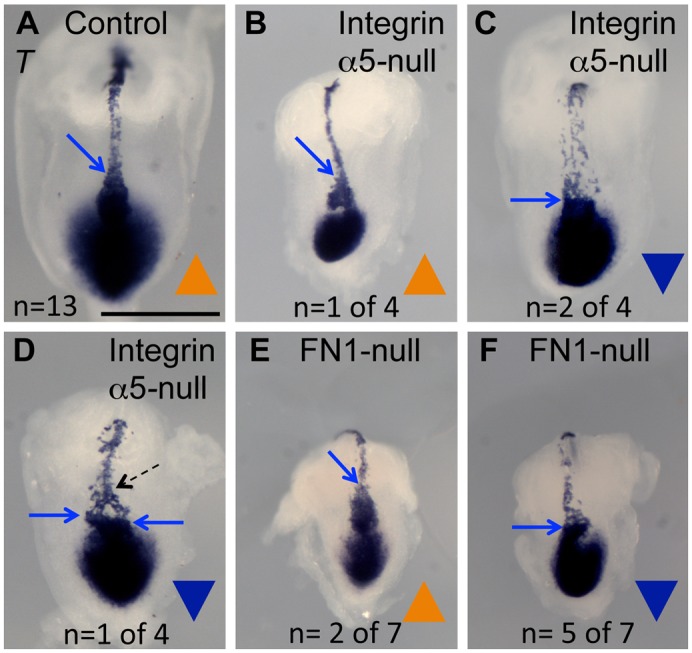
Fig. 5. Integrin α5 and FN1 regulate the geometry of the node and the development of the notochord as shown by the expression of T.

Whole-mount in situ hybridizations using an anti-sense T RNA probe. (A) Control. (B–D) Integrin α5-null mutants. (E,F) FN1-null mutants. In those mutants in which the node is oriented with the pointy end toward the anterior (blue arrows, B,E) and along the midline, the notochords are positioned at the embryonic midline and are narrow (B). In mutants with inverted nodes, notochordal cells emanate from the entire wide face of the node (blue arrow), giving rise to the wide notochord (C,F), or from two corners of the node (D). (D) Notochordal cells are refocused along the midline anteriorly (dotted arrow). Embryos were collected at E8.0. Littermate control embryos ranged from 2–5 somites. Mutants do not develop somites. Axes are as in Fig. 4. Scale bar: 500 µm.
