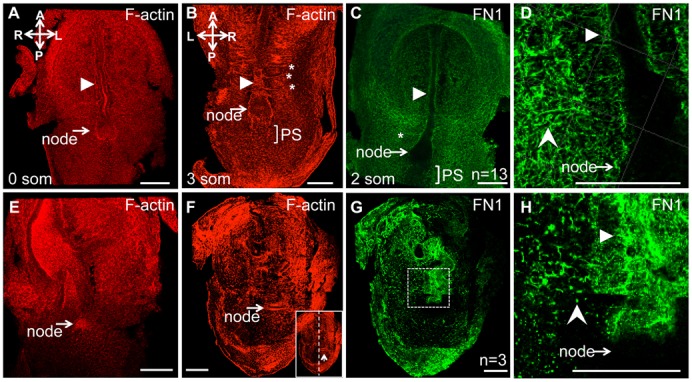
Fig. 7. Integrin α5 plays a major role in assembly of FN1 matrix, localization of F-actin, and organization of embryonic tissues.

Whole mount immunofluorescence staining of E8.0 embryos to detect F-actin (red) and FN1 (green). (A–D) Controls. (E–H) Integrin α5-null mutants. In controls, F-actin and FN1, outline the node, notochord (arrowhead) and somites (stars). In mutants, these structures are not distinguishable. Note that in the mutant (F,G) the node and the notochord are positioned to the left of the bilateral plane of symmetry. FN1 matrix is assembled as long squiggly fibrils in controls (C,D). In integrin α5-null mutants, FN1 protein is mostly present as dots (notched arrowhead, H). Filled arrowheads point at notochord, rectangle in panel G is expanded in panel H. Control and mutant embryos were isolated at E8.0. Staining for FN1 was performed on 13 control embryos ranging from 0–4 somites, and in none of the cases, FN was found in dots (notched arrowhead, H) as observed in integrin α5-null mutants.
