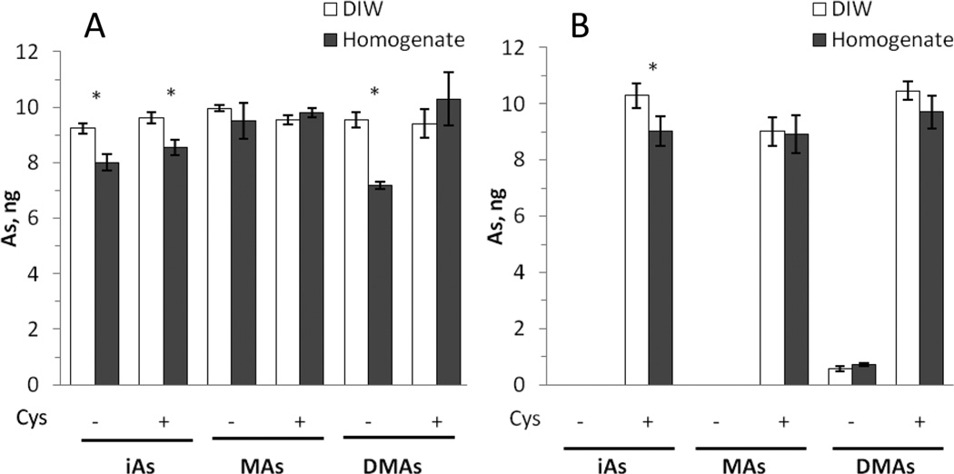
Figure 1.
HG-CT-AAS analysis of DIW and 10% liver homogenate spiked with AsIII (A) and AsV (B) standards. DIW and aliquots of the homogenate were spiked with a mixture of AsV standards (10 ng each) or with individual AsIII standards (10 ng each) and analyzed before and after pretreatment with 2% l-cysteine (Cys) (mean ± SD, n = 3). *Statistically significant differences (p < 0.05) between the amounts of an As species detected in DIW and in the homogenate as determined by ANOVA with a Bonferroni multiple comparison post-test.

