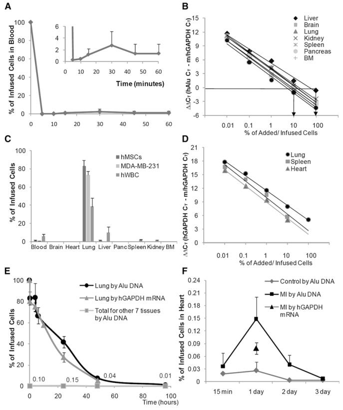
Figure 1. Assays for Fate of hMSCs Infused into Mice.
(A) Clearance of human Alu sequences from blood after i.v. infusion of about 2 × 10⁶ hMSCs into mice. Values are means ± SD; n = 6.
(B) Standard curves for real-time PCR assays of human Alu sequences in seven organs. Values indicate ΔΔCt for primers for mouse/human GAPDH genes and Alu sequences on same samples.
(C) Tissue distribution of human Alu sequences 15 min after i.v. infusion of about 2 × 10⁶ hMSCs into mice. Values are means ± SD; n = 6.
(D) Standard curves for real-time RT-PCR assays of human mRNA for GAPDH. Values indicate ΔΔCt for primers for mouse/human GAPDH genes and cDNA for human-specific GAPDH on the same samples.
(E) Kinetics of hMSCs in lung and six other tissues after i.v. infusion of about 2 × 10⁶ hMSCs. Values are means ± SD; n = 6.
(F) Appearance of hMSCs in heart after i.v. infusion of about 1 × 10⁶ hMSCs 1 day after permanent ligation of the left anterior descending coronary artery.

