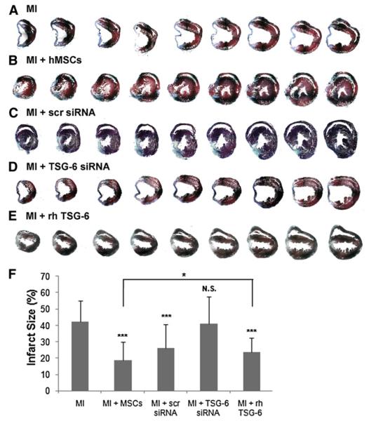
Figure 5. Assays of Infarct Size.
Each heart was cut from the apex through the base into over 400 sequential 5 μm sections and stained with Masson Trichrome. Every twentieth section is shown. Additional heart samples are shown in Figure S3.
(A–E) Symbols are as in Figure 4B, except that 100 μg rhTSG-6 protein was infused i.v. 1 hr and again 24 hr after MI.
(F) Infarct size measurements (%) obtained by midline length measurement from tenth section of the infarct area for a total of 20 sections per heart (Takagawa et al., 2007). Values are ± SD; n = 3 or 4 mice in each group; ***p < 0.0001 compared to MI controls; N.S., not significant compared to MI controls; *p < 0.05 for MI + MSCs versus MI + rhTSG-6.

