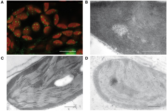
Figure 1.
Visualization of plastid nucleoids by using different microscopic techniques. (A) Nucleoids visualized by fluorescence microscopy of SYBR Green in leaf sections, bar: 10 μm. (B) Conventional electron micrographs showing nucleoids with DNA filaments in mesophyll chloroplasts. (C) Specimen prepared by high pressure freezing and freeze substitution (HPF-FS). (D) Immunogold labeling of nucleoids in leaf sections obtained from specimen prepared by high pressure freezing and freeze substitution (HPF-FS) using a DNA specific antibody, bar: 500 nm.

