
Figure 6.
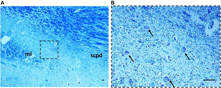
Hypoplasia of the Kölliker-Fuse nucleus in a SIUDS case (39 gestational weeks). The boxed area in (A) is represented at higher magnification in (B). At this magnification only rare suffering Kölliker-Fuse neurons are visible (see arrows). Klüver Barrera staining. Scale bar = 10 μm. ml, medial lemniscus; scpd, decussation of the superior cerebellar peduncles.