Figure 8.
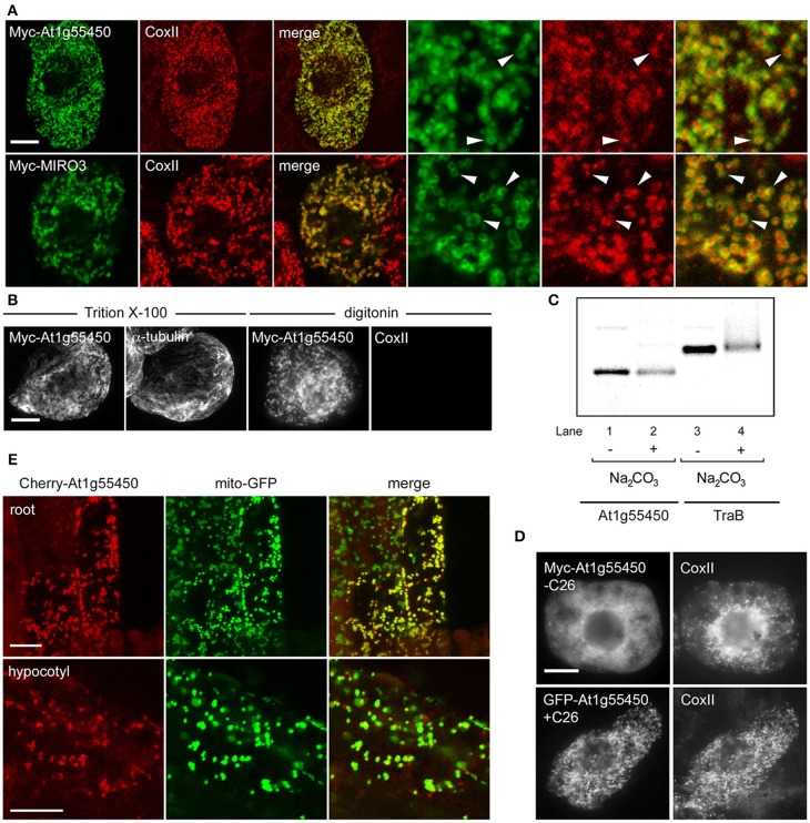
Localization, topology, membrane insertion, and C-terminal targeting-signal analysis of a novel OMM-TA protein, At1g55450. (A) Representative CLSM micrographs illustrating the localization of N-terminal Myc-tagged At1g55450 or MIRO3 to the OMM in BY-2 cells. Cells were processed for immunofluoescence CLSM as in Figure 1. Shown in the three panels on the right are images corresponding to a portion of the cell at higher magnification. Solid arrowheads indicate examples of the torus-shaped fluorescent structures containing the transiently-expressed protein delineating the spherical structures attributable to endogenous CoxII. (B) Topological mapping of Myc-At1g55450 in differential-permeabilized BY-2 cells. Cells transiently-transformed with N-terminal Myc-tagged At1g55450 were formaldehyde fixed and permeabilized with either Triton X-100 or digitonin, and then cells were processed for immuno-epifluorescence microscopy, as described in Figure 2. (C) Insertion of At1g55450 into mitochondrial membranes in vitro. Isolated pea mitochondria were incubated with in vitro synthesized Myc-At1g55450 (lanes 1 and 2) or, for comparative purposes, Myc-TraB (lanes 3 and 4), and then resuspended (+) or not (−) in alkaline Na2CO3. Equivalent amounts of each alkaline Na2CO3- or mock-extracted sample were then subjected to SDS-PAGE and phosphoimaging. (D) Representative immuno-epifluorescence micrographs illustrating the localization of a C-terminal mutant or GFP fusion of At1g55450 in BY-2 cells. Each micrograph is labeled with the name of either the transiently-expressed Myc-tagged C-terminal mutant or GFP fusion protein or endogenous CoxII. The name of each construct includes the number of amino acid residues that were either deleted from the C terminus of Myc-tagged At1g55450 (−C26) or fused to the C terminus of GFP (+C26). (E) Representative CLSM micrographs illustrating the localization of the Cherry-At1g55450 fusion protein to mitochondria in living transgenic A. thaliana seedlings co-expressing the mitochondrial marker protein mito-GFP. Labels above the panels indicate the name of the co-expressed protein and labels in panels on the left indicate the seedling tissue type. Note in the top row that not all root cells expressed the Cherry-At1g55450 fusion protein. Bars in (A and B) and (D and E) = 10 μm.

