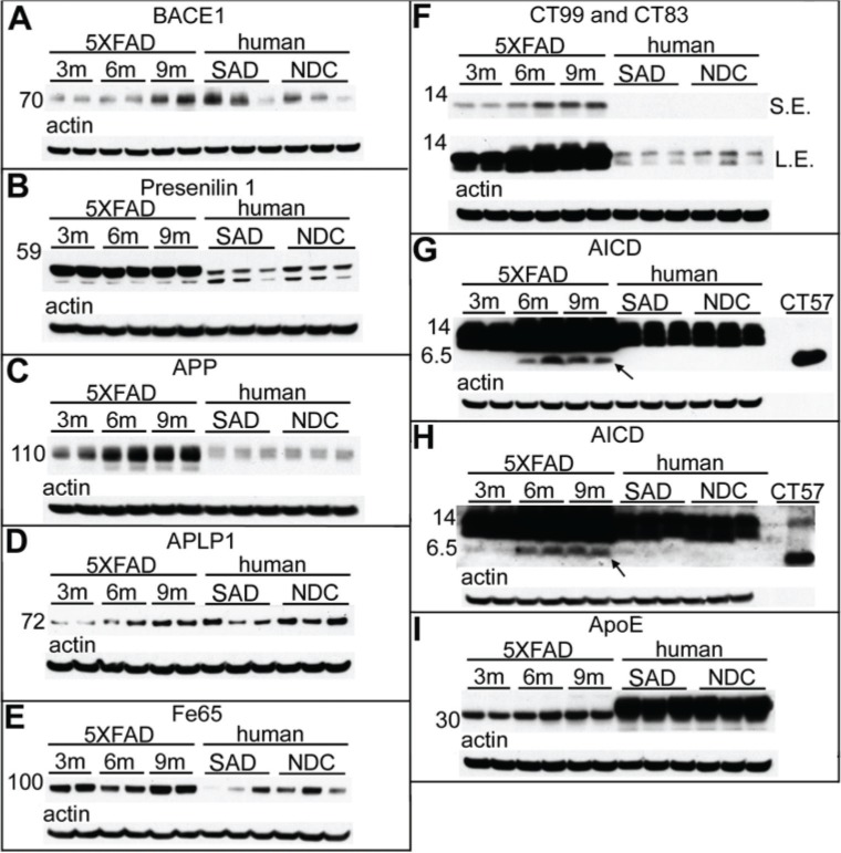
Figure 2.
Western blot analysis of male 5XFAD Tg mice at 3 months, 6 months, and 9 months of age, as well as human SAD and NDC gray matter from the frontal cortex.
Notes: (A) The mature form of BACE1 shown at ~70 kDa. (B) Full-length presenilin-1 (~55 kDa). (C) Full-length APP as detected by 22C11. (D) Full-length APLP1. (E) The adaptor protein Fe65. (F) CT99 and CT83 of APP as detected by mCT20APP. SE and LE are both shown. The putative AICD (denoted by an arrow) can be detected in older mice by antigen retrieval and by greatly increasing the antibody concentration and total protein per lane as detected with mCT20APP (G) and pCT20APP (H) antibodies. A purified CT–APP peptide of 57 amino acids (rPeptide, Athens, GA, USA) is shown on the right side of the blots in (G) and (H). (I) Full-length ApoE is observed at ~34 kDa. All blots were stripped and reprobed with actin (shown below each primary antibody) as quantitative protein loading control. A total of 40 μg of total protein was loaded into each lane for (A–F) and (I), while 65 μg of total protein was loaded into each lane for (G) and (H). For antibody details, see Table 1. The molecular weight markers are shown to the left of each blot and are reported in kDa. For further details, see Table 3.
Abbreviations: BACE, β-site APP-cleaving enzyme; APLP, amyloid-precursor-like protein; CT, C-terminal; APP, amyloid precursor protein; ApoE, apolipoprotein E; SAD, sporadic Alzheimer’s disease; NDC, nondemented controls; m, months; SE, short exposure; LE, long exposure.

