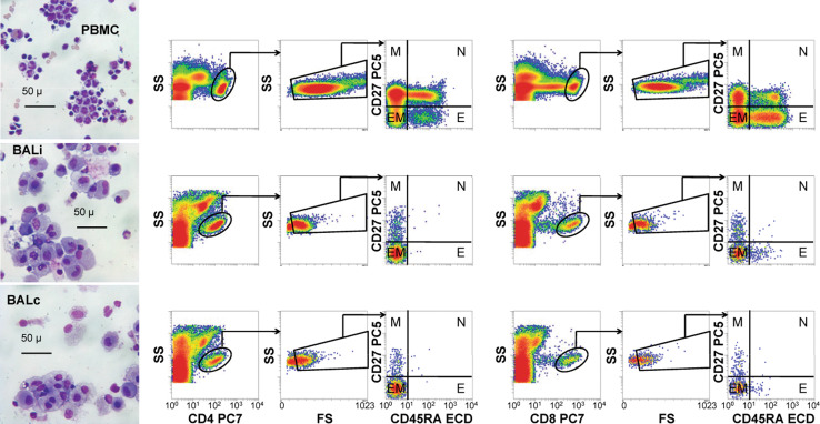
Fig. 1.
Microscopy and flow cytometry gating strategy. Wright–Giemsa-stained cytocentrifuge preparations were photographed using a ×40 objective (left column). The images were viewed to assess the general quality and contents of each sample before flow cytometric analysis. The flow gating strategy for these samples involved the selection of either CD4+ or CD8+ events, elimination of low light scatter (apoptotic) events, and classification into memory (M), naïve (N), effector/memory (EM), or effector/terminally differentiated (E) subsets (middle and right panels). Patient ipsilateral bronchoalveolar lavages (BALi), patient contralateral bronchoalveolar lavages (BALc), patient peripheral blood mononuclear cells (PBMC), and normal PBMCs (not shown) were analyzed in this way

