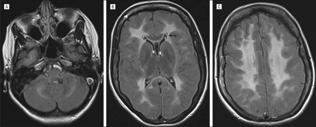
Figure 1.
Axial fluid-attenuated inversion recovery magnetic resonance images. A, Symmetric involvement is noted at the level of the cerebellar white matter. B, Diffuse and symmetric leukodystrophy is evident; the U fibers, the outer rim of the corpus callosum, and the internal capsule are relatively spared. Small areas of cystic degeneration (arrow) are noted bilaterally in the periventricular white matter. The typical widespread white matter rarefaction is lacking. C, Diffuse and symmetric white matter involvement is noted at the level of the centrum semiovale.

