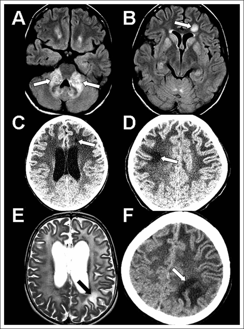
Figure 1.
Focal white matter lesions in 3 Alexander disease cases. Multiple imaging studies showing focal lesions in case 1 (A, B), case 2 (C, D), and case 3 (E, F). A–B, noncontrast axial FLAIR images at age 13 showing bright lesions with mass effect in the middle cerebellar peduncles (A) and a left frontal focal well-defined lesion (B). C–D, noncontrast axial CT images: first, at age 6 years, there is a small left frontal hypodense lesion (C), and then by age 9 years there is good visualization of an additional right superior frontal hypodense lesion (D). E–F, axial T2 MRI (E) and noncontrast axial CT image (F) showing some left parietal white matter that is more bright in signal than is the surrounding abnormal white matter at age 6 years (E) and has distinctively abnormal appearance in comparison to surrounding white matter on CT at age 7 years (F). CT, computed tomography; FLAIR, fluid-attenuated inversion recovery; MRI, magnetic resonance imaging.

