
Figure 1.
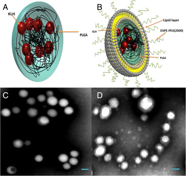
Schematic illustration and TEM images of the NPs. (A) Schematic illustration of PK NPs. (B) Schematic illustration of LPK NPs. (C) TEM image of PK NPs, which highlights the uniform size and spherical shape of PK NPs. (D) TEM image of hybrid LPK NPs, which shows the lipid-bilayer-enclosed PK NPs. The scale bars represent 200 nm.