
Figure 2.
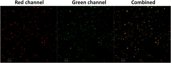
Confocal images of LPK NPs. The images illustrate that KLH was labeled with rhodamine B (red) and liposome was labeled with NBD (green), confirming that PK NPs were enclosed by liposome. Scale bars represent 10 μm.
Official websites use .gov
A
.gov website belongs to an official
government organization in the United States.
Secure .gov websites use HTTPS
A lock (
) or https:// means you've safely
connected to the .gov website. Share sensitive
information only on official, secure websites.
Confocal images of LPK NPs. The images illustrate that KLH was labeled with rhodamine B (red) and liposome was labeled with NBD (green), confirming that PK NPs were enclosed by liposome. Scale bars represent 10 μm.