
Figure 6.
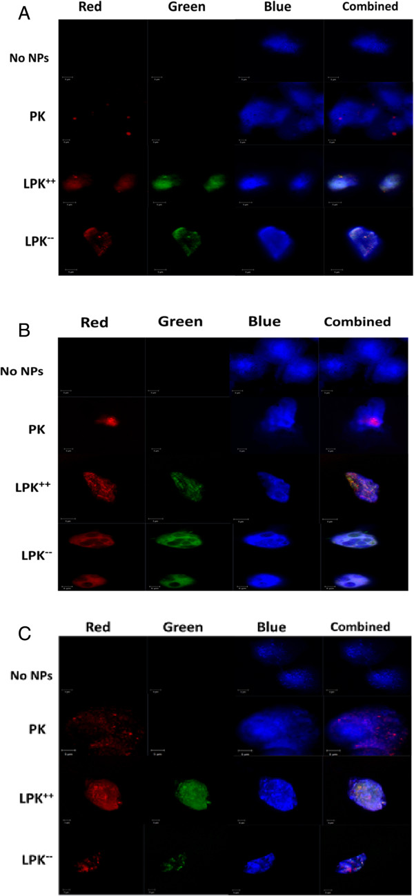
Confocal images of internalization of PK NPs and LPK NPs by JAWSII DCs. One hundred thousand cells were incubated with 0.1 mg NPs for 1 h (A), 2 h (B), and 3 h (C), respectively. The incubation concentration was 0.2 mg/mL. Red color is from rhodamine B, which was used to label KLH; green color is from NBD PE, which is a fluorescent lipid used to label the lipid layer; and blue color is from CellMask™ Blue Stain, which was used to label the cell membrane. Both positively charged LPK NPs and negatively charged LPK NPs were internalized more readily by cells than PK NPs. Scale bars represent 5 μm.