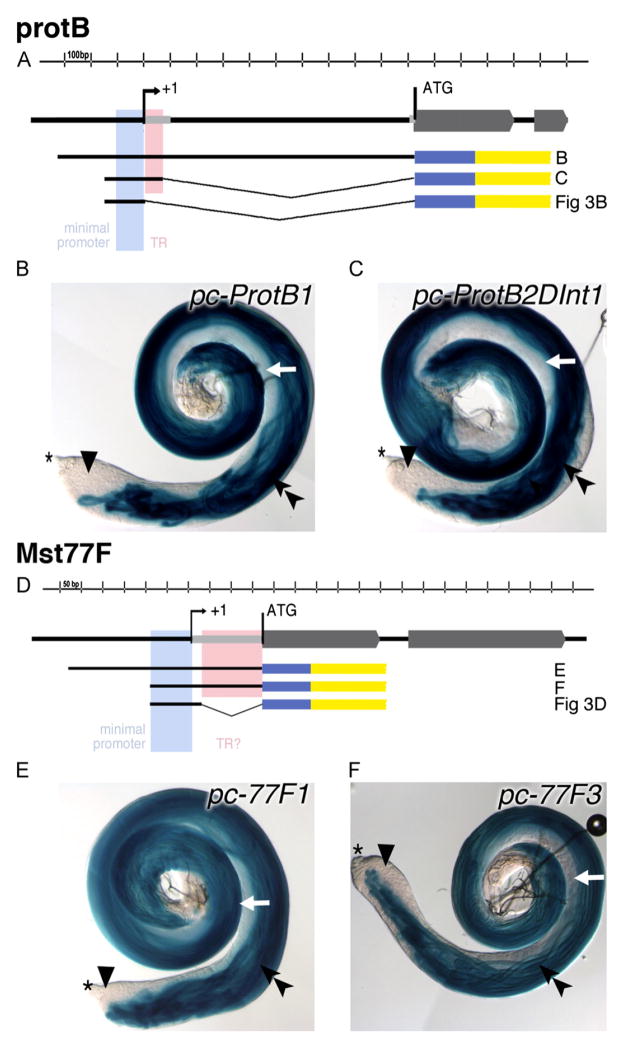
Fig. 2.
Expression of ProtB and Mst77F is controlled by short upstream regions. (A, D) Schematic drawings of the genomic regions and the generated promoter-lacZ constructs of protB and Mst77F. Exons are depicted as gray block arrows, the minimal promoters are in light blue, and the regions tested for contributing to translational control (TR) are in pink. Black thick lines illustrate the regions contained within each promoter-lacZ construct; thin gray lines indicate the deleted regions. The reporter gene lacZ is indicated in blue; the SV40 3′ UTR with a polyadenylation signal is indicated in yellow. (B, C, E, F) Analyses of β-Galactosidase activity in the testis of transgenic flies bearing different protB (B, C) or Mst77F (E, F) promoter lacZ constructs after 10 min of staining reaction. Asterisk indicate the tip of the testis. Double arrowheads, elongated spermatid bundles; arrowheads, spermatocytes; and early spermatids, white arrows.

