Fig. 6.
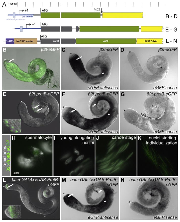
The ORF of protB confers translation in late spermatids. (A) Diagram of the constructs. (B, E, H–L) eGFP fluorescence; (C, D, F, G, M, N) in-situ hybridization using an eGFP-specific probe. (B, C, D) eGFP under the control of the β2-tubulin promoter and 5′ UTR (β2t-eGFP). (B) Whole mount of adult testis; arrow, late spermatids. (C) Hybridization with an eGFP antisense probe; arrow, early spermatocytes; arrowhead, early spermatids. (D) Hybridization with an eGFP sense probe. (E, F, G) protB-eGFP under the control of the β2-tubulin upstream control region and 5′ UTR (β2t-proB-eGFP). (E) Whole mount of adult testis; arrows, late spermatids; inset, higher magnification. (F) Hybridization with an eGFP antisense probe; arrow, early spermatocytes, arrowhead, early spermatids; double arrowhead, indicates late signal in spermatids. (G) Hybridization with an eGFP sense probe. (H, I, J, K) Squash preparations of β2-protB-eGFP testes, GFP autofluorescence and α-histone, (H) Spermatocytes, (I) Young elongating nuclei, (J) Canoe-stage nuclei. (K) Nuclei starting individualization (L, M, N) bam-GAL4-driven UAS-ProtB-eGFP. (L) Whole mount of adult testis; Arrow, spermatogonia; inset, higher magnification of the tip of this testis. (M) Hybridization with an eGFP antisense probe; arrow, spermatogonia; arrowhead, spermatid stage; double arrow, eGFP transcripts hardly detectable distally to this point. (N) Hybridization with an eGFP sense probe; double arrow, spermatids. In (B–G, L–M), the asterisk marks the hub region of the testis.

