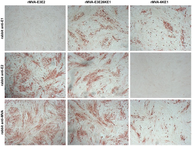
Figure 2. Detection of antigen expression.
BHK-21 cells were infected with the respective candidate MVA vaccines and 24 hours later cells were fixed in methanol/acetone (1∶1) and stained for specific expression of CHIKV E1 (upper panel), E2 (middle panel) or MVA antigens (lower panel). The respective antigens were detected using rabbit polyclonal serum specific against E1, E2, and MVA as indicated. The staining confirmed the specific expression of the respective antigens and their purity. *Images were contrast enhanced in Adobe Photoshop.

