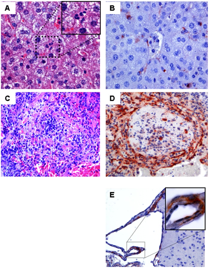
Figure 4. Histopathology of several tissues staining positive for CHIKV antigen.
Panels A–E show representative staining of the liver, spleen, and brain of CHIKV infected AG129 mice. Hematoxylin and eosin-stained sections of the liver from all groups showed no abnormalities (A; 40× objective). Endothelial and Kupffer cells stained positive with anti-CHIKV capsid antibody (B, 40× objective). Massive depletion of lymphocytes was observed in the spleen of animals that died after challenge with CHIKV (C; HE staining, 40× objective). Antigen was mainly located in endothelial cells in the spleen (D). Epithelia cells of the choroid plexus (E) in the brain were scarcely stained and no antigen was found in the neuropil of the brain. Examples of positively stained cells are indicated by block arrows.

