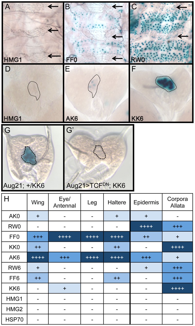
Figure 5. Tissue-specific activity of HMG-Helper pair reporters.
Brightfield images of tissues from 3rd instar larva containing the indicated lacZ reporter constructs stained for lacZ activity. (A–C) LacZ expression in larval epidermis. Expression is seen in cells underlying the naked cuticle, located between denticle belts (arrows). The HMG1 reporter has no detectable expression; FF0 has weak expression and RW0 drives robust expression. (D–F) Ring glands with expression in the Corpora Allata (dotted lines). HMG1 has no detectable expression with AK6 and KK6 displaying weak and strong expression, respectively. (G–G′) Expression of TCFDN in the CA (via the Aug21Gal4 driver) abolishes expression of the KK6 reporter. (H) Summary of expression data from all tissues examined, with number of plus signs and blue hue indicative of the relative level of reporter gene expression. At least 12 samples of each reporter line were analyzed for each tissue, with similar results observed between individual samples.

