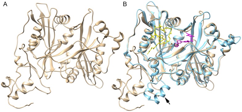
Figure 3. Modeling the structure of B. malayi NMT and structural comparison with Leishmania major NMT.
(A) Ribbon diagram of the predicted crystal structure of NMT from B. malayi. (B) Comparison of B. malayi NMT (tan) with L. major NMT (blue: 2wsa). An overlay of the structure of these enzymes using UCSF Chimera [26] reveals a nearly identical conformation of the binding sites for myristoyl-CoA (yellow) and inhibitor DDD85646 (magenta). The 2 small helixes (arrow) formed by an insertion of 21 amino acids in L. major NMT are replaced with a loop in B. malayi NMT.

