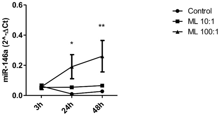
Figure 1. MiR-146a expression in cells exposed to M. leprae.
Macrophage-like THP-1 cells (5×10⁵) were infected with live M. leprae (MOI 10∶1, 100∶1) for 3, 24 and 48 h at 33°C. RNA was extracted and a real-time stem-loop RT-PCR was performed using RNU48 to normalize. Data show mean ± SEM (*p<0.05 relative to 24 h control, **p<0.05 relative to 48 h control). Results represent four independent experiments.

