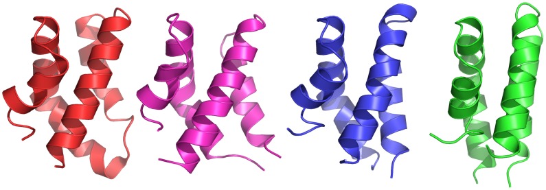
Figure 8. Comparison of folded potato PSI to other SAPLIPs.

Structural comparison of the folded potato PSI at pH 4.5 (blue) and pH 7.4 (green), averaged over the last 200 ns of the simulation trajectories, to the crystal structure of barley PSI (PDB ID 1QDM, magenta) and the crystal structure Sap C (PDB ID 2GTG, red). Potato PSI simulated at pH 7.4, simulated with parameters closely resembling the experimental parameters used for both barley PSI and Sap C, exhibited a compact globular structure consisting of a distorted four-α-helix bundle characteristic of other SAPLIPs. Potato PSI simulated at pH 4.5 adopted a compact four-α-helix bundle structure not previously observed for any SAPLIP. The linker regions of potato PSI are omitted for clarity.
