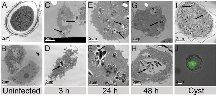
Figure 4. Transmission electron microscopy showing uninfected (A) cysts and (B) trophozoites as well as the intravacuolar location of M. ulcerans (arrows) within infected, intact trophozoites (C, D) 3 h, (E, F) 24 h, (G, H) 48 h and (I) cysts 3 weeks post infection.
(J) is a composite image showing a cyst 22 days post infection by fluorescence microscopy.

