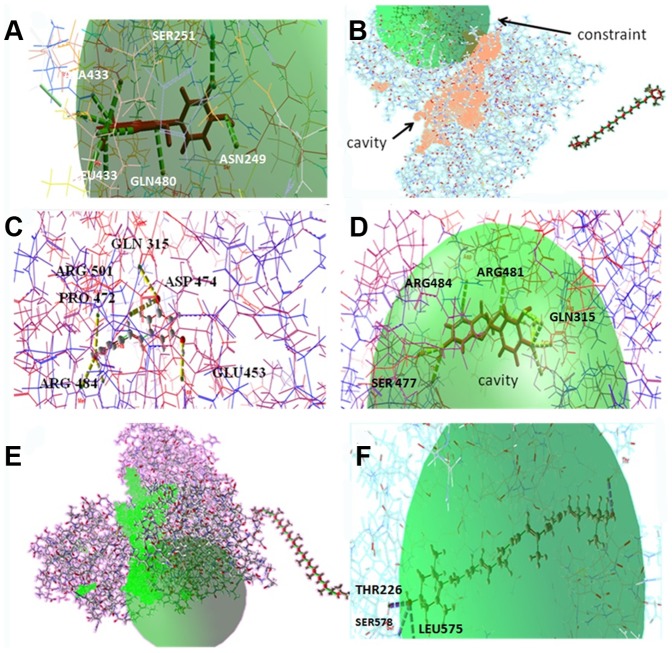
Figure 7. Docking conformations and binding pockets of HCVGT3a NS3 helicase with different inhibitors.
(A) Three-dimensional representation of quercetin with target macromolecule and its hit residues (B) Beta-carotene represents no interactions in its constraints with the modeled structure. The Hbond formation is shown in stick mode (green). (C) Best pose of the compound resveratrol forming seven HBonds with three-dimensional structure of helicase at its binding sites GLN 315, GLU 453, ARG 484, PRO 472, ASP 474, ARG 501 (D) Three-dimensional representation of catechins and target molecule with eleven HBonds network. The Hbond formation is shown in stick mode (yellow) and the constraint is shown in green color. (E) Zero interactions between lycopene and modeled GT3a NS3 helicase (F) binding of lutein at THR 226, LEU 575 and SER 578 residues. Lutein forming three HBonds with high HBond energy −2.5461 kcal·mol−1.

