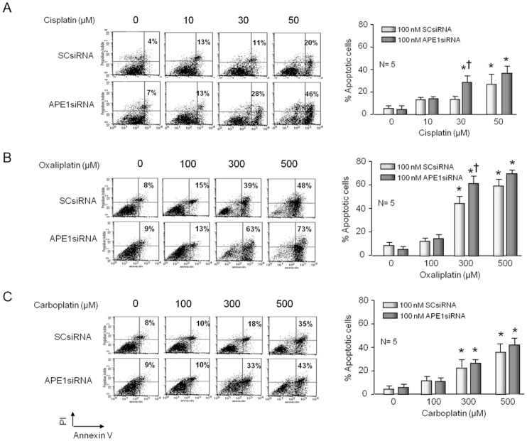
Figure 2. Apoptosis induced by cisplatin and oxaliplatin, but not carboplatin, is increased by reducing the expression of APE1 in sensory neuronal cultures.
Neuronal cultures were treated with siRNAs on days 3–5 in culture then exposed to various concentrations of platins for 72 hours starting on day nine in culture. Cell apoptosis was detected by Annexin-V and PI staining and FACS analyses after cells were grown for 12 days. The left panels show representative fluorescence-activated cell sorting (FACS) for cells treated with various concentrations of cisplatin (A), oxaliplatin (B), or carboplatin (C) and scrambled siRNA (SCsiRNA) or APE1siRNA as indicated. The panels on the right show the quantification of data from five independent harvests. Each column represents the mean ± SEM of the percent of apoptotic cells from cultures treated with SCsiRNA (lightly shaded) or with APE1siRNA (heavy shaded) and various concentrations of cisplatin (A); oxaliplatin (B) or carboplatin (C) as indicated. An asterisk indicates significant difference in survival in the absence or presence of drug treatment, whereas a cross indicates significant difference in cultures treated with SCsiRNA versus APE1siRNA using Student's t-test.

