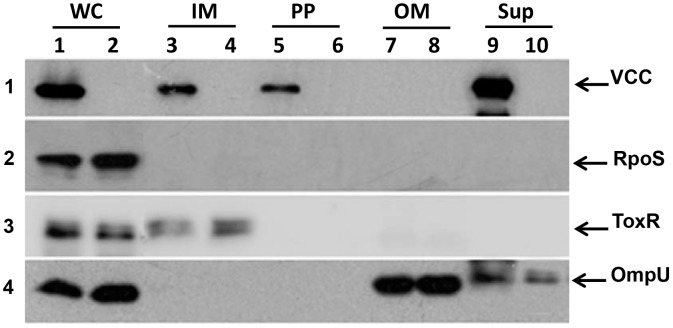
Figure 1. Immunoblot analysis of cell fractions.
(A) Whole cell lysate (WC), Inner membrane (IM), periplasmic (PP), outer membrane (OM) fractions and culture supernatant (Sup) from the NOVC strain V:5/04 (lanes 1, 3, 5, 7, 9) and its isogenic Δvcc mutant (MDS-1) (lanes 2, 4, 6, 8, 10), respectively. The samples were subjected to immunoblot analysis to detect VCC and different internal controls to rule out cross-contamination during fractionation; RpoS (as a cytoplasmic marker, panel 2), ToxR (as a inner membrane marker, panel 3), and OmpU (as an outer membrane marker, panel 4).

