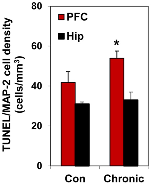
Figure 3. PFC is more vulnerable to ethanol-induced neuronal apoptosis than hippocampus.

Brain sections double-labeled with TUNEL and MAP-2 in the PFC and hippocampus (Hip) and quantified by stereological counting; Values are means ± SEM; *p<0.01. Neither the number of MAP-2-positive cells (neurons) nor volumes of the brain structures were affected by chronic 3-week ethanol exposure. Note significantly higher density of TUNEL- positive neurons in PFC than in hippocampus of ethanol-exposed mice.
