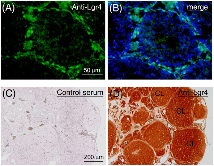
Figure 5. Distribution of the Lgr4 protein in the postnatal gonads.
To assess the testicular distribution of Lgr4, testicular sections from mice at 5 weeks old were probed with the anti-Lgr4 antibody followed by fluorescein isothiocyanate-conjugated secondary antibody. Data are shown in parallel without (A) or with (B) a DAPI merged image. To assess the ovarian distribution of Lgr4, ovarian sections from mature rats (8 weeks old) were probed either (C) with preimmune rabbit serum or (D) with the anti-Lgr4 antibody. The sections were then visualized using a horseradish peroxidase system. CL, corpus luteum.

