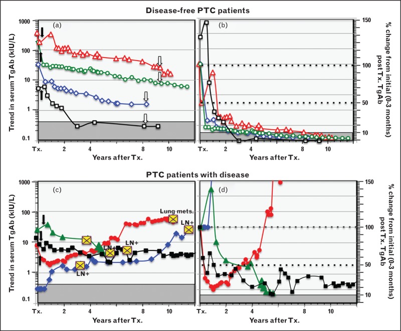
FIGURE 4.

Thyroglobulin autoantibody trend and percentage change used as a surrogate differentiated thyroid cancer tumor-marker. This figure illustrates how the trend in TgAb concentrations (left panels a and c) and the percentage change in TgAb concentrations (Kronus/RSR method) relative to an initial (0–3 month) postoperative TgAb value (panels b and d) can be used as a surrogate DTC tumor-marker. Figure 4a shows TgAb trends for four PTC patients who were judged disease-free by ultrasound (open arrows) in the postoperative period. Figure 4b shows these data converted to percentage of the initial value. Disease-free patients may have a transient early TgAb rise in response to thyroglobulin released by surgical injury or RAI treatment (solid arrows), but thereafter TgAb values typically decline over time (years) to less than 10% of initial level. These disease-free patients were selected to have different initial TgAb concentrations to illustrate that declining TgAb concentrations do not necessarily become undetectable during follow-up, unless the initial TgAb was low. Figures 4c and 4d show comparative data for four PTC patients with persistent or recurrent disease detected during follow-up (indicated by yellow crosses). A de-novo TgAb appearance, a TgAb rise or a stable TgAb concentration that fails to fall below 10% of initial value are indications of active disease. DTC, differentiated thyroid cancer; LN, lymph node metastases; PTC, papillary histotype; RAI, radioiodine; TgAb, thyroglobulin autoantibody; Tx., thyroidectomy.
