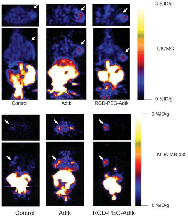
FIGURE 4.
microPET of tumor-bearing mice (top: subcutaneous U87MG tumor on right shoulder; bottom: orthotopic MDA-MB-435 tumor on left mammary fat pad) 1 h after injection of 18F-FHBG (10-min static scan). Images (both trans-axial and coronal) were normalized to the same scale. The tumors are indicated by white arrows in all cases.

