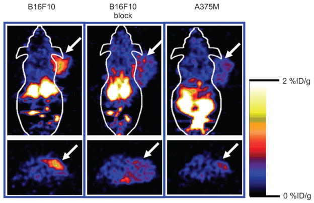
FIGURE 4.
Decay-corrected coronal (top) and transaxial (bottom) microPET images of C57BL/6 mouse bearing B16/ F10 tumor (left and middle images) and Fox Chase Scid mouse bearing A375M tumor (right images). Images were acquired 1 h after tail vein injection of 18F-FB-NAPamide (3.7 MBq [100 μCi]) with or without coinjection of 200 μg NDP peptide. Arrows indicate location of tumors.

