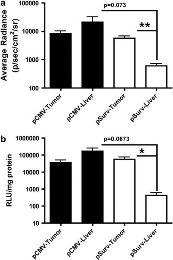
Figure 5.
Analysis of BLI imaging and luminometer measurements of tissue lysates. (a) Analysis of cut tumor images revealed a nonsignificant trend towards increased FL activity in the normal liver compared with the tumor in rats infected with Ad-pCMV-FL (mean±s.e.m.; n=3). In contrast, the Survivin-targeted virus (n=4) significantly improved the tumor specificity of transgene expression, and showed a strong trend towards abrogating FL activity in the normal liver compared with the CMV-targeted virus. Importantly, administration of either the Survivin- or CMV-targeted viruses resulted in similar levels of FL activity within tumors. (b) Luminometer readings of FL activity from tumor or normal liver lysates from animals infected with either virus corroborated the imaging results. *P<0.05 and **P<0.01 as determined via a two-tailed, unpaired t-tests.

