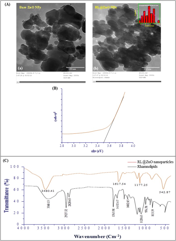
Figure 5. Characterization of RL@ZnO nanoparticles.
(A) The representative TEM microphotographs of bare ZnO nanoparticles (a) and RL@ZnO nanoparticles (b) at an accelerating voltage of ∼200 kV. Inset of the figure (b) depicts the particle size analysis. (B) Band gap plot of RL@ZnO nanoparticles, calculated by Tauc plot formula. (C) FTIR spectra of RL extract (red line) and RL@ZnO nanoparticles (black line). The spectra shown are representatives of the three independent experiments.

