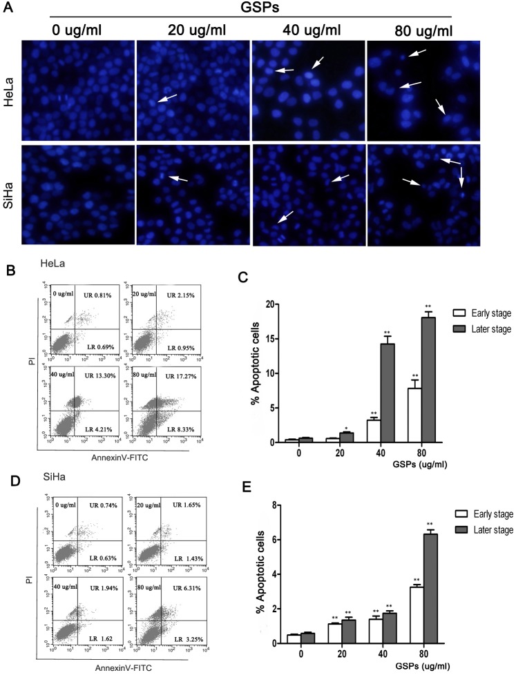
Figure 3. GSPs induced the apoptosis of cervical cancer cells in vitro.
HeLa and SiHa cells were treated with varying doses of GSPs for 48 h and then harvested for analysis of apoptosis. (A) The morphological changes of nuclei were examined by fluorescence microscopy using DAPI staining. The arrow indicates nuclear condensation and an apoptotic body (magnification, 40×). Flow cytometry analysis of Annexin V-FITC/PI double-stained HeLa (B) and SiHa (C) cells. The low right (LR) quadrant of the histograms indicates the early apoptotic cells, and the upper right (UR) quadrant indicates the late apoptotic cells. The treatment of HeLa (B) and SiHa (C) cells with GSPs results in significant increases in the percentages of apoptotic cells (including early stage and late stage). Values are expressed as the mean ± SD of three experiments in duplicate, *p<0.05 vs. control; **p<0.01 vs. control.

