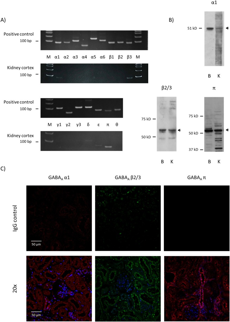
Figure 1. GABAA receptor subunits are expressed in rat kidney.
A) RT-PCR results for GABAA receptor subunits in rat (WKY on normal diet) kidney cortex. Appropriately-sized bands were detected for GABAA receptor α1, β3 and π subunits in at least five independent experiments. Rat brain RNA or commercial universal RNA was used as positive control. M: molecular marker. B) Immunoblot for GABAA receptor α1, β3 and π subunits in rat kidney cortex. B: brain control, K: kidney cortex. C) Immunofluorescent examination of GABAA receptor α1, β3 and π subunits in rat kidney cortex. Staining with normal IgG instead of primary antibody served as negative control.

