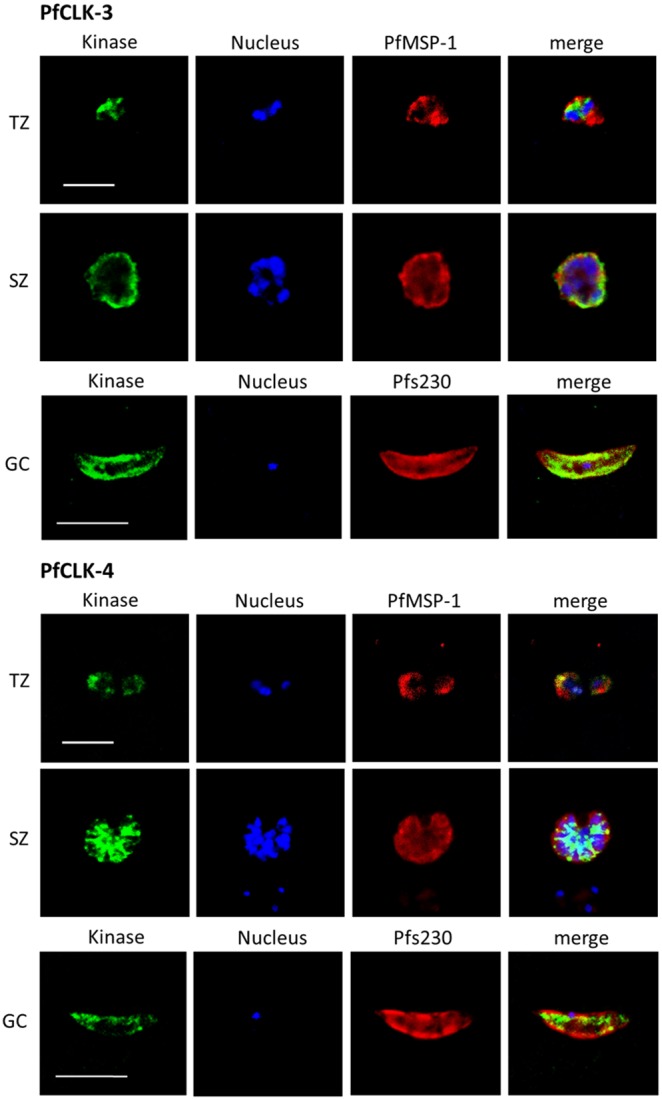
Figure 3. Subcellular localization of PfCLK-3 and PfCLK-4 in the blood and gametocyte stages.

Mixed asexual blood stage cultures containing trophozoites (TZ) and schizonts (SZ) and mature gametocyte (GC) cultures were fixed with methanol and prepared for IFA, using rat antisera against PfCLK-3 and mouse antisera against PfCLK-4 (green). The parasite nuclei were highlighted by Hoechst staining (blue). The asexual blood stages were labelled with rabbit antisera against PfMSP-1 and gametocytes with rabbit antisera against Pfs230 (red). Bar, 5 µm.
