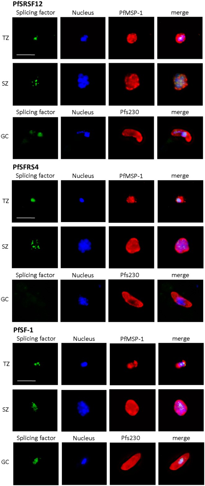
Figure 5. Subcellular localization of the SR proteins in the blood and gametocyte stages.

Mixed asexual blood stage cultures containing trophozoites (TZ) and schizonts (SZ) and mature gametocyte (GC) cultures were fixed with paraformaldehyde and prepared for IFA, using mouse antisera against PfSRSF12, PfSFRS4 and PfSF-1 (green). The parasite nuclei were highlighted by Hoechst staining (blue). The asexual blood stages were labelled with rabbit antisera against PfMSP-1 and gametocytes with rabbit antisera against Pfs230 (red). Bar, 5 µm.
