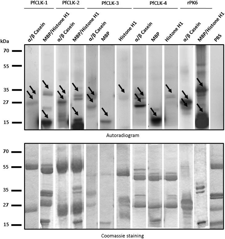
Figure 7. Phosphorylation of exogenous substrates by immunoprecipitated PfCLKs.
Kinase activity assays were deployed to detect phosphorylation of the substrates histone H1, MBP and α/β casein (∼33, 18, and 28/34 kDa, respectively; indicated by arrows) by the four immunoprecipitated PfCLKs (autoradiogram, upper panel), using the PfCLK-specific respective mouse, rabbit or rat antisera. Assays without precipitated proteins (PBS control) were used for negative controls. Recombinant protein kinase 6 (rPK6) was used for positive control. Coomassie blue staining (lower panel) of radiolabelled SDS-gels was used as a loading control.

