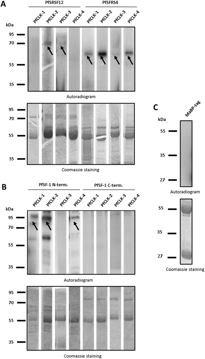
Figure 8. Phosphorylation of plasmodial SR proteins by immunoprecipitated PfCLKs.
A. Kinase activity assays were deployed to detect phosphorylation of recombinant PfSRSF12 and PfSFRS4 (∼73 and 65 kDa, respectively; indicated by arrows) by two or more of the PfCLKs (autoradiogram; upper panel). B. The N-terminal part (∼95 kDa; indicated by arrows), but not the C-terminal part (86 kDa) of recombinant PfSF-1 was phosphorylated by immunoprecipitated PfCLKs. An additional phosphorylation signal of truncated N-terminal PfSF-1 was visible at approximately 60 kDa. C. MaBP-tag alone (43 kDa) as substrate was used as negative control. Shown here is an assay using PfCLK-3-specific immunoprecipitate, similar results were obtained with immunoprecipitates of other PfCLKs (not shown). Coomassie blue staining (lower panels) of radiolabelled SDS gels was used as a loading control.

