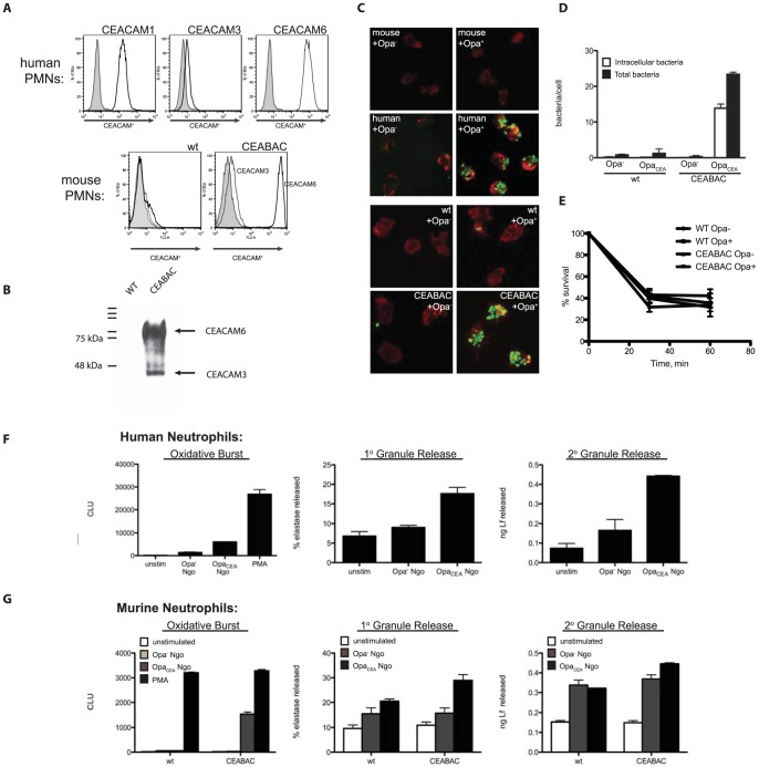
Figure 1. Human CEACAM expression by mouse neutrophils results in neisserial capture and internalization.
(A) Human CEACAMs are expressed in CEABAC neutrophils in a manner reflecting that in human neutrophils. Human neutrophils (top), or wild type (WT) and CEABAC mouse neutrophils (bottom) were fixed and stained with antibodies specific for CEACAM1, CEACAM3, CEACAM6, or a mouse IgG isotype control, and analyzed by flow cytometry. Isotype histograms are shaded. (B) Immunoblot showing CEACAM expression in WT and CEABAC neutrophils. (C) Mouse neutrophils do not bind N. gonorrhoeae, while human neutrophils bind N. gonorrhoeae in an Opa-dependent manner. Mouse (top) or human (bottom) neutrophils were infected with non-opaque (Opa−) or Opa-expressing (Opa+) N. gonorrhoeae. Cells were visualized by staining filamentous actin with phalloidin [16], and bacteria are shown in green. Intracellular and total bacteria were differentially stained, and quantified via immunofluorescence microscopy (D). (E) WT and CEABAC PMNs kill Opa− and Opa+ bacteria with similar kinetics. Adherent WT and CEABAC PMNs were infected with either Opa− or Opa+ N. gonorrhoeae at an MOI = 1. Bacterial survival over time was evaluated as CFUs present in PMN lysates at each time point relative to bacterial CFUs present at time 0. N = 2. (F–G) N. gonorrhoeae infected CEABAC neutrophils respond analogously to human PMNs. Human (F) or mouse (WT and CEABAC) (G) neutrophils were infected with Opa− or Opa+ N. gonorrhoeae and oxidative burst and degranulation responses were analyzed as described in Materials and Methods.

