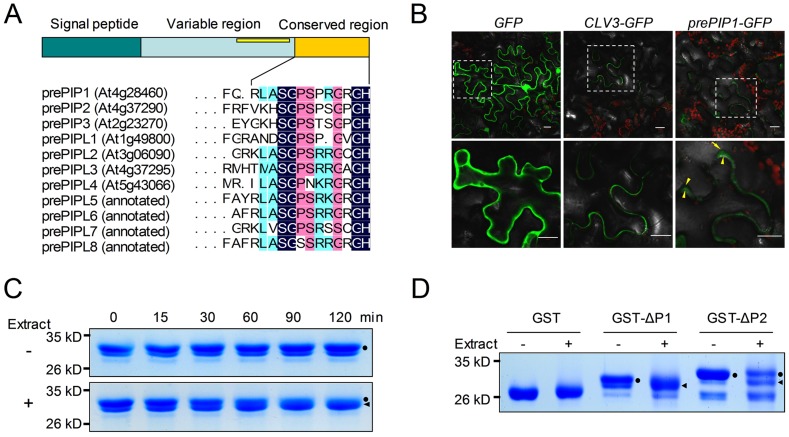
Figure 1. Identification of PIP peptides.
(A) Schematic presentation of prePIP homologs in A. thaliana. (B) Sub-cellular distribution of prePIP1-GFP in tobacco leaf cells. Tobacco leaves were transformed with Agrobacterium GV3101 harboring a construct containing GFP, prePIP1-GFP or CLV3-GFP, respectively. The yellow arrows point the plasma member. Scale bar = 20 µm. (C) Time-course of GST-ΔP1 proteolytic processing. (D) Proteolytic cleavage of GST-ΔP1 and GST-ΔP2 by total protein extract from A. thaliana. (C–D) SDS-PAGE separation of protein products. Dots mark intact GST-ΔP1 or GST-ΔP2; triangles mark processed GST-ΔP1 or GST-ΔP2. At least three replicates were performed with similar results.

