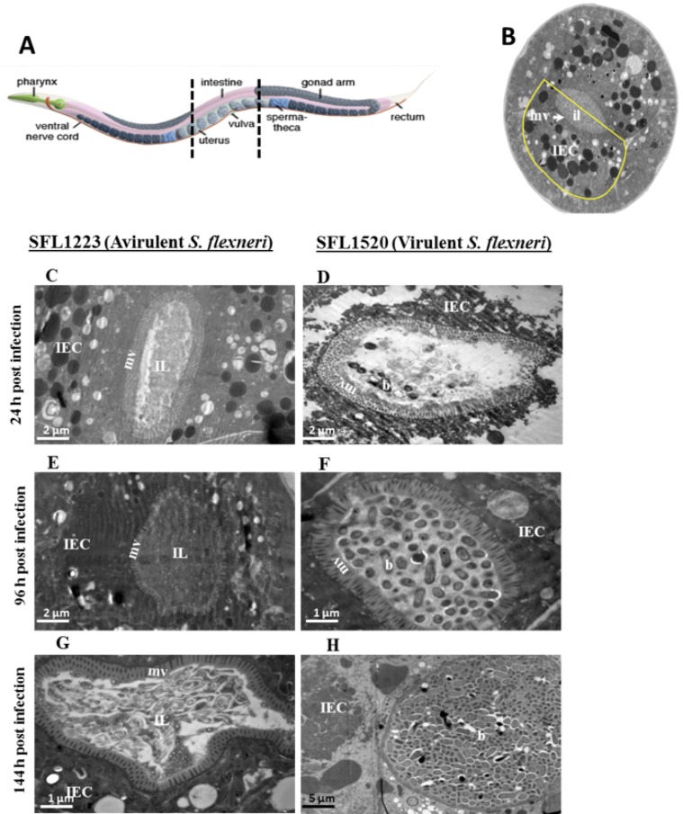
Figure 3. Virulent S. flexneri cells escape pharyngeal grinding and accumulate in the C. elegans intestinal lumen.
A: Schematic representation of the C. elegans body plan with the plane of sections highlighted (artwork by Altun and Hall, © Wormatlas). B: Transverse section of the mid body of a healthy nematode with the intestinal cell highlighted in yellow. C–H: Transmission electron microscopy micrographs of transverse mid body sections of animals feeding on plasmid-cured, avirulent S. flexneri (SFL1223) (C, E, G) and virulent S. flexneri serotype 3b (SFL1520) (D, F, H) for 24 h (C, D), 96 h (E, F) and 144 h (G, H). IEC-intestinal epithelial cell; mv-microvilli; IL-intestinal lumen; b-intact S. flexneri cells.

