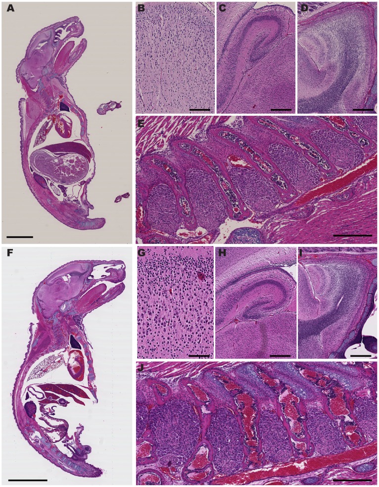
Figure 2. Normal anatomy of Nav1.7 KOs.
Postnatal day 4 Nav1.7 WT (A to E) and KO (F to J) neonates stained with hematoxylin and eosin (H&E). Wild type: A. Sagittal section of the entire pup. B through E show magnifications of various regions of the central and peripheral nervous systems: Knock Out: F. Sagittal section of the entire pup. G through J show magnifications of various regions of the central and peripheral nervous systems. (B, G) cortex; (C, H) hippocampus; (D, I) olfactory bulb; (E, J) dorsal root ganglia. (Scale bars: A, F = 5 mm; B, G = 100 µm; C, H and D, I = 400 µm; E, J = 300 µm).

