. 2014 Aug 28;8:459. doi: 10.3332/ecancer.2014.459

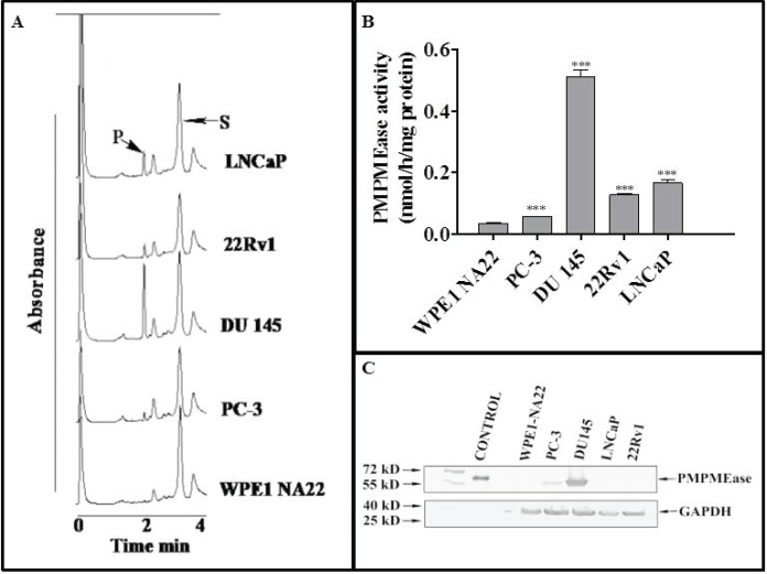
Figure 1. PMPMEase is hyperactive in prostate cancer cells. Cell lysates (1.3–2.8 mg of protein) were incubated with RD-PNB substrate (1 mM) at 37°C and the reaction mixtures were analysed by HPLC as described in the methods. Panel A shows HPLC chromatograms for the analysis of samples from the different prostate cells for PMPMEase hydrolysis of the substrate, while Panel B shows the computed specific PMPMEase activities in the cell lysates. Panel C shows the relative PMPMEase protein levels determined by Western blotting when compared to the pure PMPMEase (3.3 µg, positive control) and normal prostate cells by paired t-test. ***p < 0.0001.

Figure 1.