
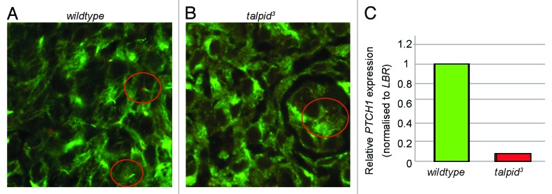
Figure 4. Hedgehog signaling is perturbed in the talpid3 liver during development. IHC anti-acetylated (cilia axonemes, green) and γtubulin (centrosomes, red) E6 (A andB). Wt liver has cilia axonemes projecting from centrosomes (circled) (A) cilia axonemes are not observed projecting from centrosomes in the talpid3 liver (circled) (B). (C) Real-time PCR identified a 0.08-fold reduction in PTCH1 expression in the day 6 talpid3 liver, compared with the wt.