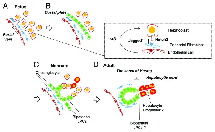
Figure 1. Liver development and transition of tissue localization of LPCs. (A) Hepatoblasts are fetal bipotential liver stem/progenitor cells (LPCs), which abundantly exist in fetal liver by mid gestation. Around E15, hepatoblasts near the portal vein are committed to cholangiocytes by the activation of Notch signaling pathways through direct interaction with Jagged-1+ portal fibroblasts as well as TGFβ by receiving the ligand secreted from endothelial cells and/or fibroblasts. (B) The ductal plates are the primitive structure of bile ducts. Cholangiocytes in this structure function as LPCs, which have the ability to differentiate into hepatocytes. (C) In late gestation and neonatal period, cholangiocytes establish tubular structures though part of the cells still exist in the ductal plate. During this period, many cholangiocytes maintain the ability to differentiate into hepatocytes and may function as LPCs. (D) During postnatal development, most of the cholangiocytes lost the ability to differentiate into hepatocytes. However, a small number of LPCs exist in normal adult liver. Although their tissue localization has not been definitely identified, LPCs may exist in or near the canal of Hering, the boundary between hepatic cord and bile ducts.

An official website of the United States government
Here's how you know
Official websites use .gov
A
.gov website belongs to an official
government organization in the United States.
Secure .gov websites use HTTPS
A lock (
) or https:// means you've safely
connected to the .gov website. Share sensitive
information only on official, secure websites.
