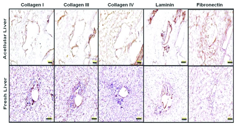
Figure 1. ECM protein localization on native human liver scaffold and native human liver. Immunostaining for collagen I, III, IV, fibronectin, and laminin (as indicated) show similar ECM component distribution in prepared acellular liver bioscaffold and native human liver sections. Scale 50μm (From Hepatology with permission from Wiley and Sons).

An official website of the United States government
Here's how you know
Official websites use .gov
A
.gov website belongs to an official
government organization in the United States.
Secure .gov websites use HTTPS
A lock (
) or https:// means you've safely
connected to the .gov website. Share sensitive
information only on official, secure websites.
