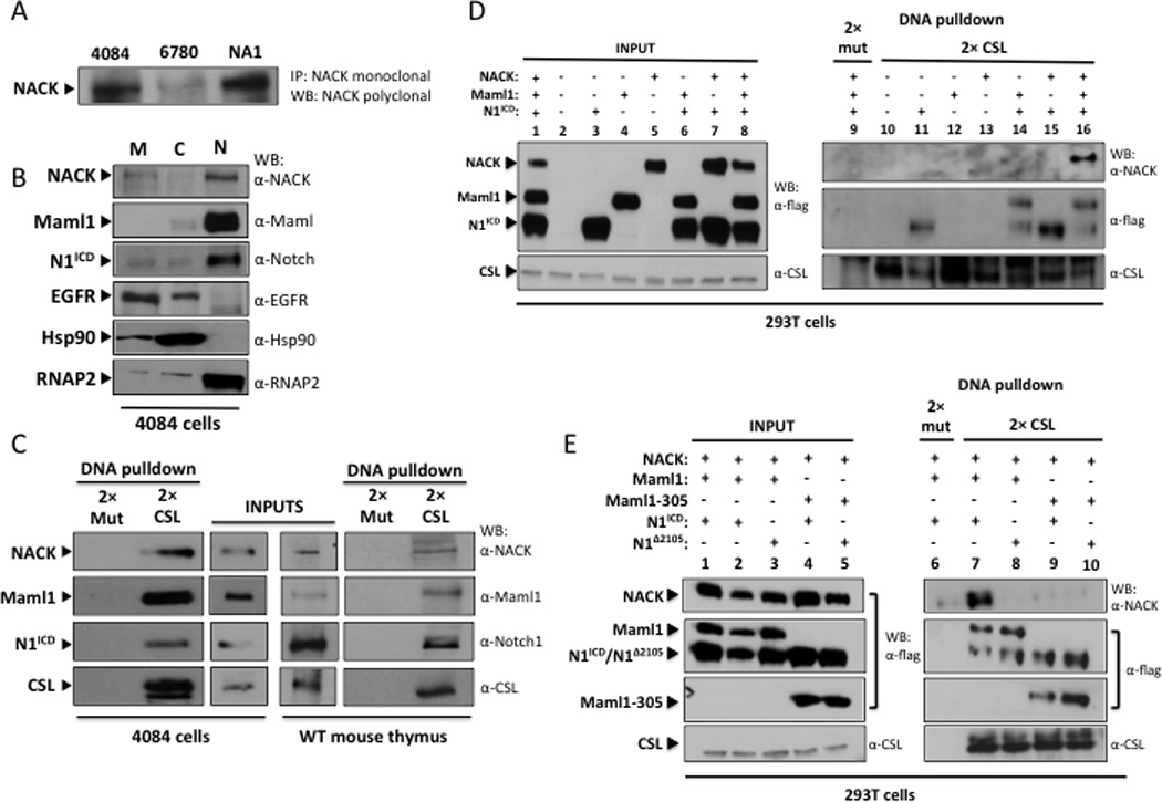
Figure 1. NACK forms a complex with N1ICD and Maml1.

A. IP/western analysis of lysates from T-ALL cell lines. 4084 – Notch-induced lymphoma; 6780 – Myc-induced lymphoma; NA-1 – Ikaros-null lymphoma. B. Western analysis of subcellular fractions of 4084 cells. M – membrane; C – cytoplasm; N – nuclear. C. DNA pull-down and western analysis of nuclear lysates from 4084 cells and wt mouse thymocytes using beads conjugated to 2× CSL binding DNA or 2× mutant CSL binding DNA (2× mut). Input lanes represent 5% of total nuclear lysate. D. DNA pull-down and western analysis of lysates from 293T cells transfected with different combinations of N1ICD, Maml1, and NACK. Input lanes represent 10% of total lysate. E. DNA pull-down and western analysis of lysates from 293T cells transfected with different combinations of N1ICD, N1Δ2105, Maml1, Maml1–305, and NACK. Input lanes represent 10% of total lysate.
