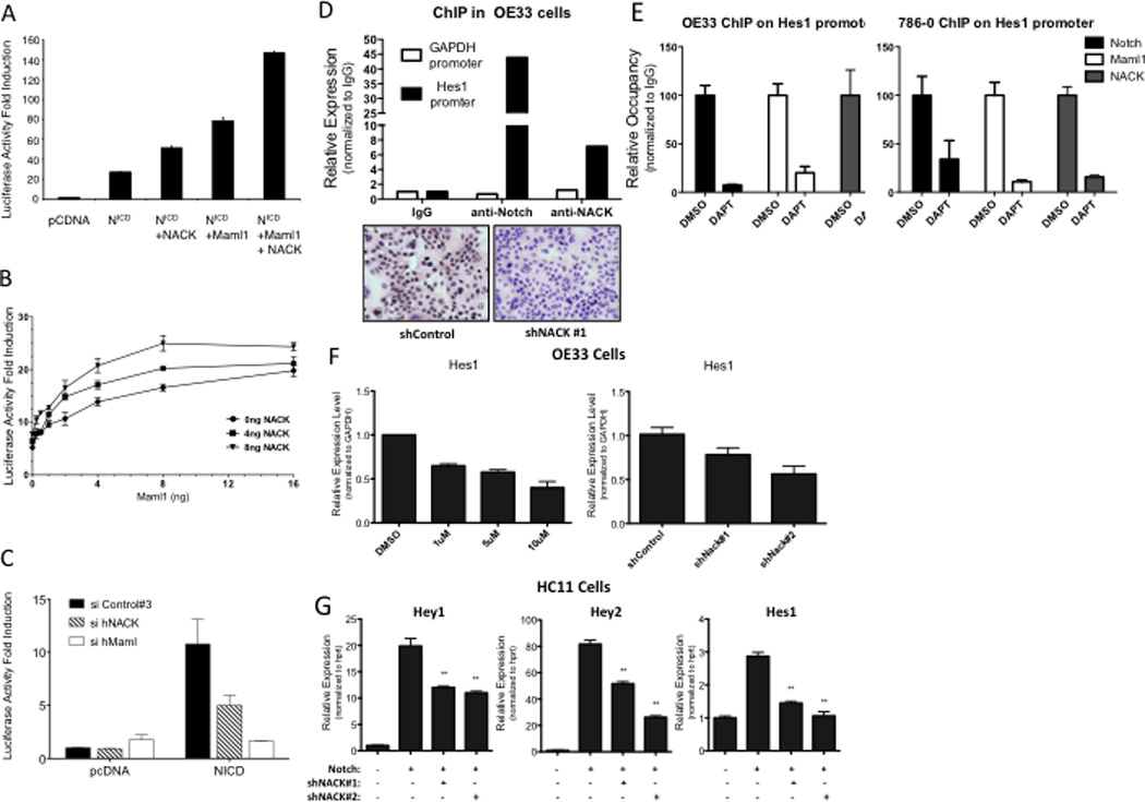
Figure 2. NACK is a co-activator of Notch signaling.

A–C. 8× CSL luciferase reporter assays in H1299 cells transfected with different combinations of plasmids. Experiments were performed in triplicate and bars represent mean (SEM). A. Histogram of luciferase activity. B. Titration curve showing luciferase activity with different quantities of Maml1 and/or NACK over a fixed quantity of N1ICD (2 ng). C. Histogram of luciferase activity after siRNA-mediated knockdown of Maml or NACK. D. ChIP of Notch and NACK on the Hes1 promoter in OE33 cells. IHC validates specificity of the α-NACK antibody. E. ChIP of Notch, Maml, and NACK on the Hes1 promoter in OE33 and 786-0 cells after treatment with DAPT. F. Hes1 expression in OE33 cells treated with DMSO or DAPT, or infected with shRNA against NACK. Bars represent mean (SEM) of 3 samples. **p<0.01 versus shControl. G. Expression of Notch target genes in HC11 cells infected with N1ICD and shRNA against NACK. Bars represent mean (SEM) of 3 samples. **p<0.01 versus Notch.
